Chest Imaging

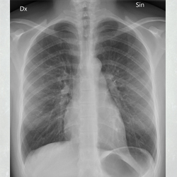
A CXR is produced by exposing the chest to a small dose of radiation called ‘ionising radiation’.
- Different parts of the body absorb different amounts of the x-rays which is how we see the different parts of the body on the image showing up from black, shades of grey to white. Bones absorb the x-rays and appear white on the CXR.
- Normal air filled lungs let the x-rays pass through so appear black.


