Invasive tests - Bronchoscopy
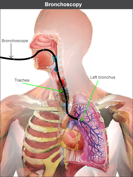
Bronchoscopy is a test where a fibre optic telescope with a camera is inserted into the nose or mouth, is passed through the vocal cords and down into the airways that supply the right and left lung.

A bronchoscopy is performed for many reasons:
- To visualise and biopsy abnormal or cancerous areas seen on a CT scan.
- To test for lung infections.
- To look for abnormalities not seen on a CT scan if there are undiagnosed and persistent symptoms, for example, cough or coughing up blood.
- To take a lung biopsy ( a small lung tissue sample) to diagnose lung conditions, particularly in interstitial lung disease which has many types and causes.
During the bronchoscopy
- Patients will need to have fasted (no food or drink for a few hours) before the procedure.
- Oxygen levels, heart rate and blood pressure will be monitored during the bronchoscopy.
- A sedative is given intravenously (in to a vein). This will not make the patient unconscious, but it should make them feel more relaxed.
- A local anaesthetic is given to the back of the throat, over the vocal cords and over the airways to reduce coughing.
- A camera is inserted in to the upper airway and the images from the camera are seen on a small screen allowing the operator to direct the scope inside the body.
- The procedure is usually over within 5-10 minutes.


